
SIMULATING REALITY <em>The Value of Simulated Use Models in MedTech Development</em>

PART 1: UNDERSTANDING THE PHYSIOLOGICAL ENVIRONMENT
September 16, 2026
Annette Branger, PhD
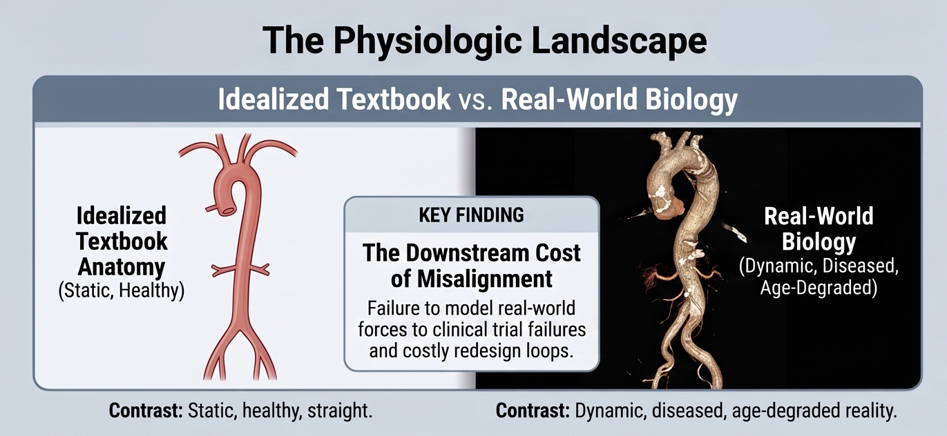
One of the most challenging aspects of designing a medical device is developing a deep understanding of the physiologic environment and the range of forces imparted on the device, during its use. This multi-part article series will discuss some considerations when making a simulated use model intended to replicate the physiologic conditions for testing of medical devices used in vivo, either acutely or as a permanent implant, to ensure their design is sufficiently robust for the expected application. Too often, the literature, textbooks, and other available sources do not adequately represent the diseased state, the appropriate patient demographics, and the inherent range of motion and forces created by these conditions. Getting the physiologic conditions wrong has far-reaching downstream implications, potentially causing unintended and unwanted patient consequences and disrupting clinical trials for device redesigns. Understanding the physiologic environment and creating a realistic Simulated Use or anatomical model to replicate those forces, is therefore crucial to medical device design and development.
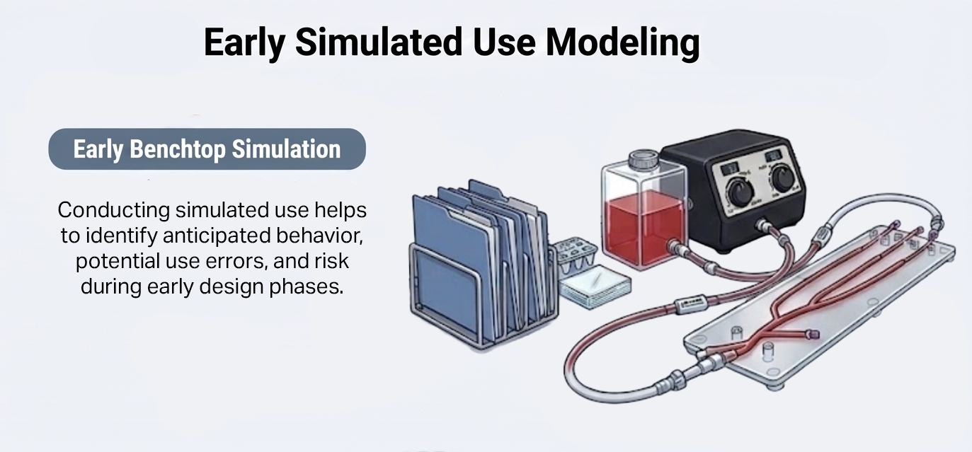
Creating this model early in the design and development process helps identify device shortcomings or unexpected behavior in a timely way. When used in Design Verification and Validation testing, this Simulated Use model can highlight the robustness of the device design and identify any use errors that may arise. Successful execution of Simulated Use testing can also greatly minimize the risk of having to manage device failures in the field or in clinical trials.
So how do you properly understand the physiologic environment, both from a geometric and a motion perspective, in order to replicate it on the benchtop? First, if the device is not completely novel, there may be FDA or other regulatory guidances outlining crucial testing parameters. These regulatory documents, however, are sometimes frustratingly vague, asking the medical device manufacturer to justify the boundary conditions with little guidance on what they should be.
As you dig into the literature, it's also worth commenting on both the utility and the shortcomings of AI, now that it has become such a common research tool. AI is a great jumping-off point, giving the user threads of resources and literature to pull on; however, it often misquotes or misunderstands the literature it cites and the data it presents. The motto “trust but verify” applies to AI, which should never be relied on at face value. The original source that AI drew from should always be reviewed, and what you find might surprise you or lead you to other valuable information. AI can also serve as a useful second check, identifying information you may have missed.
In these cases, resources aside from published literature may include higher-resolution imaging or 3D reconstructions from the appropriate patient groups. There are many open-source, public domain, and paid databases that contain patient images for all parts of the anatomy. Even better, if applicable, are databases containing dynamic imaging, such as MRI or ultrasound, showing anatomical structures in motion.
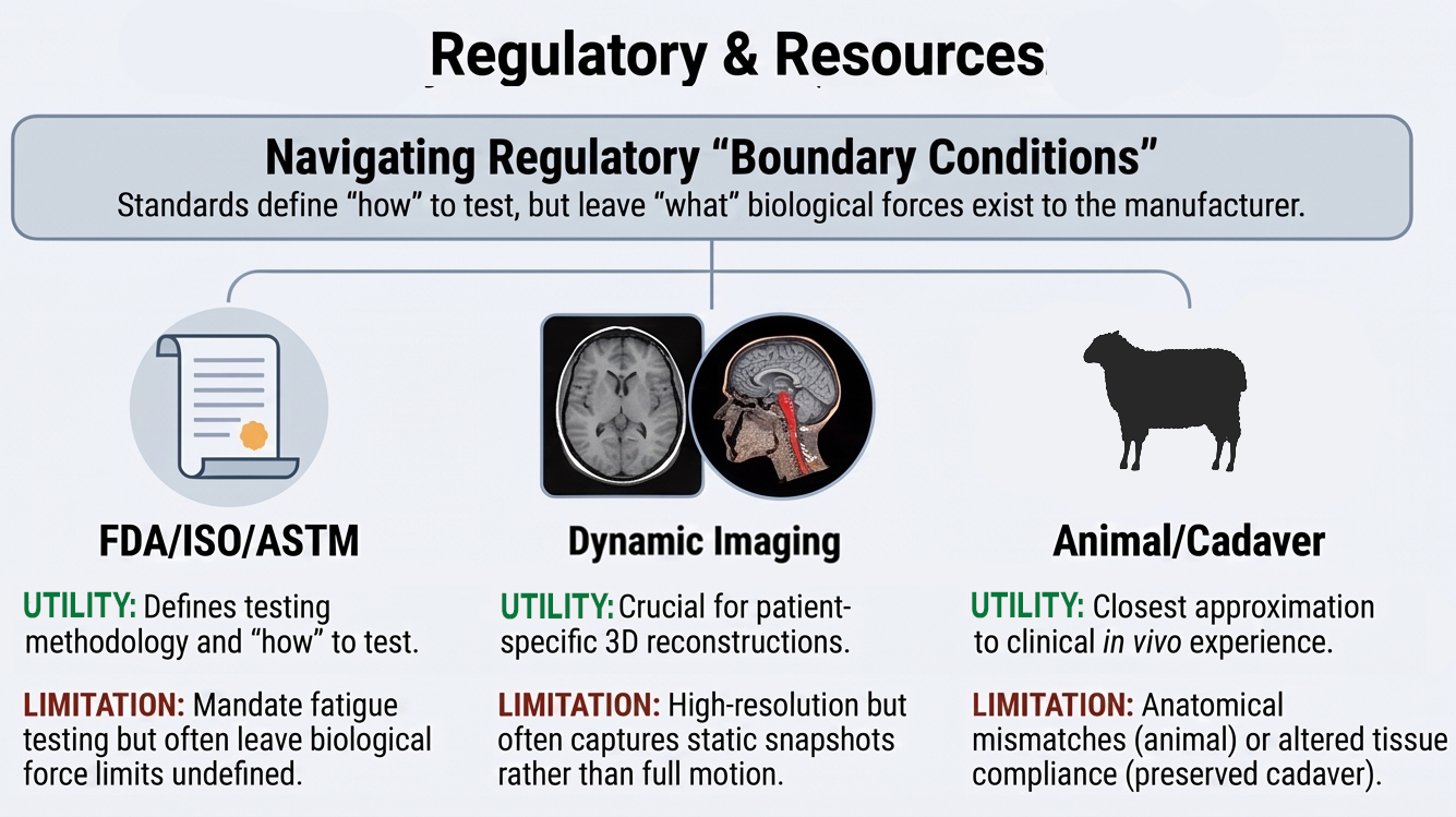
Another valuable resource for gaining a proper understanding of the physiologic environment is animal models or cadaveric models. Using the right animal species for the right application, to test and analyze device behavior in vivo through various imaging modalities, can provide a scenario as close to the clinical experience as possible. These studies, however, can be costly, and they come with real limitations: there may be obvious anatomical differences between species, and the native tissue used is typically in a healthy state rather than a diseased one, so the absence of an actual disease state can affect results.
Cadaver tissue, alternatively, can provide anatomically accurate measurements with limited movement, though it comes with its own set of drawbacks. Pressurizing vessels to physiologic levels and replicating true physiological blood flow are difficult to reproduce in cadaveric tissue, and the preserved, non-living tissue also degrades and loses the elasticity of native vessels, changing how it behaves compared to living anatomy. With each of these resources, it is important to consider and account for their limitations when using the data as input for your own benchtop Simulated Use model.
Two cautionary tales illustrate what happens when the physiologic environment is underestimated, even in seemingly relatively simple anatomy, and the problems it can create for medical device manufacturers. The first involves early stents used in the Superficial Femoral Artery (SFA) for atherosclerotic disease. Textbook images of the SFA present the artery as a relatively straight vessel (Figure 1); in practice, however, implanted metallic stents were often found to have catastrophic fractures. Following a period of intense scrutiny and research, analysis of the vessel in a variety of patient positions has showed that the SFA does not maintain that textbook straight configuration, but instead twists, bends, and stretches, imparting unaccounted-for forces on the stents.² The limb flexion portrayed in Figure 1 demonstrates that flexion transforms the artery from a relatively straight conduit into a shorter, curved, compressed, and twisted pathway. Deformation is greatest near the adductor hiatus and in the popliteal artery behind the knee. A device must accommodate this changing anatomical state without kinking, lumen compromise, fracture, migration, or loss of apposition.
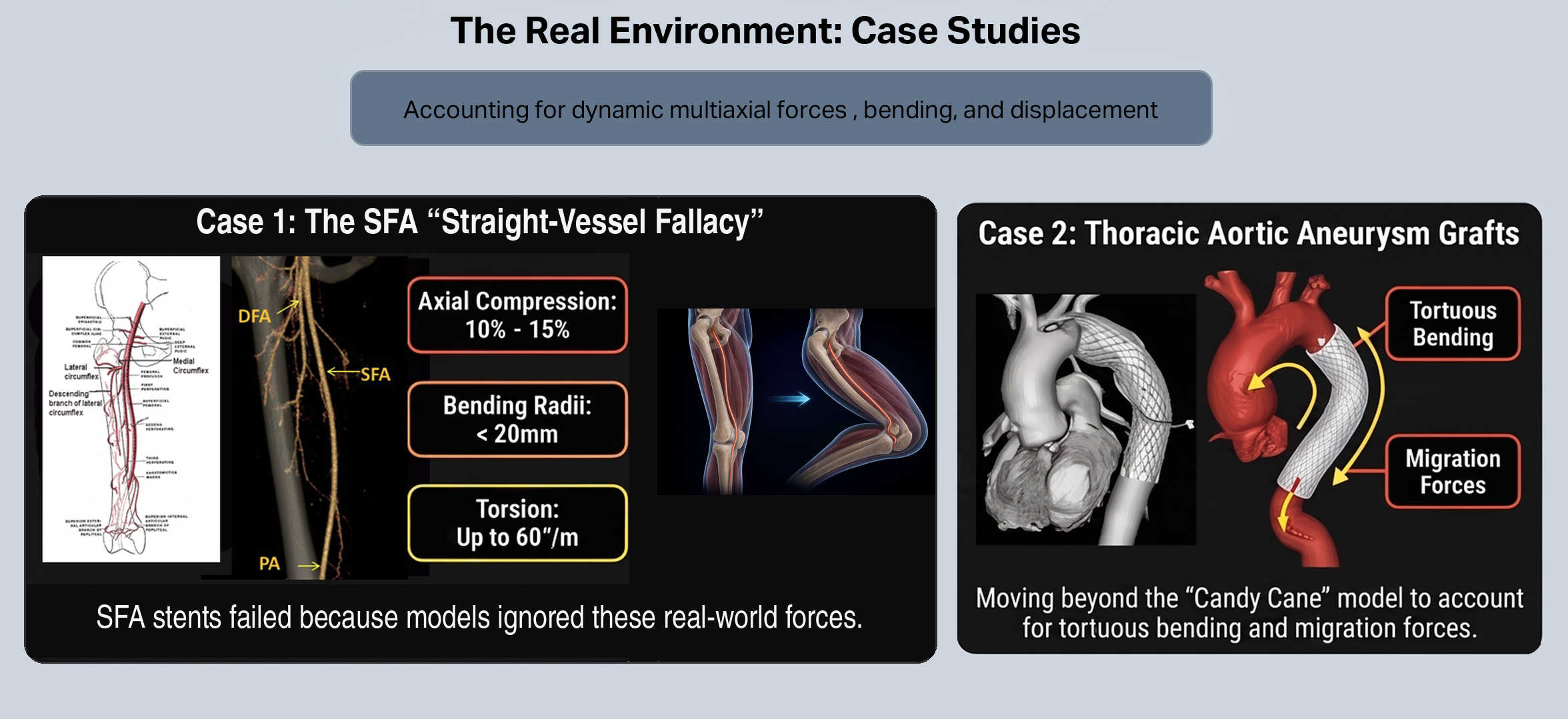
Figure 1. Case 1: The superficial femoral artery as commonly depicted in textbooks compared to CT Angiography for evaluating arterial anatomy¹’³ and dynamic anatomy under motion²
Figure 2. Case 2: Aortic Tortuosity and Motion impact on device behavior and integrity
A similar story played out in the early days of thoracic aortic aneurysm stent grafts, when the thoracic aortic anatomy was always depicted as a straight “candy cane” shape (Figure 2). After early failures, device manufacturers realized that the patient population presenting with thoracic aneurysms often had anatomies that were anything but straight.⁴ These tortuous aortas had motion that went well beyond simple radial compliance, with significant concomitant bending. Once these anatomies and motions were understood, manufacturers, while still facing real engineering challenges, were able to produce devices that could withstand these environments.
Both cases point to the same lesson: the environment a device will actually live in rarely matches the one drawn in a textbook, and the gap between the two is where failures happen. Part 2 of this article series turns to the next step, translating that understanding of the physiologic environment into an actual benchtop Simulated Use model.
References:
Khan A, Arain A. Anatomy, Bony Pelvis and Lower Limb: Anterior Thigh Muscles. In: StatPearls. StatPearls Publishing; 2023. Updated March 17, 2023. Accessed September 13, 2026. https://www.ncbi.nlm.nih.gov/books/NBK538425/
Sousa Pereira, J. A., Tinto, H. A. M. R., Fernandes, L., Bilhim, T., & Seabra, Z. (2013, March 7–11). CT Angiography and Digital Subtraction Angiography in the evaluation of atherosclerotic peripheral arterial disease – pictorial essay [Poster presentation]. European Congress of Radiology (ECR 2013), Vienna, Austria. https://doi.org/10.1594/ecr2013/C-1638
Shahbad R, Pipinos M, Jadidi M, et al. Structural and mechanical properties of human superficial femoral and popliteal arteries. Ann Biomed Eng. 2024;52(4):794-815. doi:10.1007/s10439-023-03435-3
Belvroy VM, Romarowski RM, van Bakel TMJ, et al. Impact of aortic tortuosity on displacement forces in descending thoracic aortic aneurysms. Eur J Vasc Endovasc Surg. 2020;59(4):557-564. doi:10.1016/j.ejvs.2019.09.022